
Foot X Rays – How Does It Work & What You Can Expect?
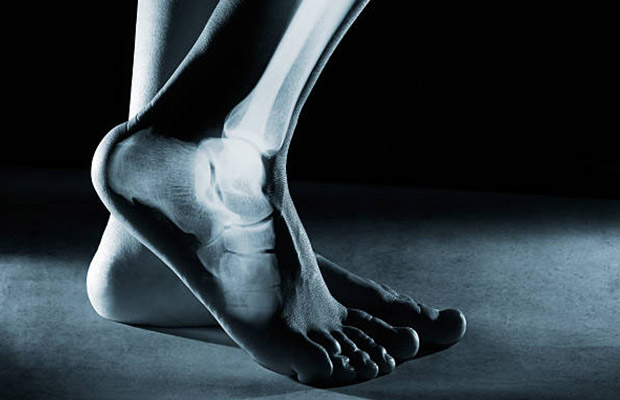
A foot X-ray is a test that produces an image of the anatomy of your foot. Your healthcare provider may use foot X-rays to diagnose and treat health conditions in your foot or feet.
Foot X-rays are a simple, quick, and painless process. Your leg will be positioned on an X-ray table by a radiologic technician who will then take numerous images of it.
Everything you need to know about foot X-rays is included in this post, keep reading!
Table of Contents
What Are X-rays?
Radiation known as electromagnetic waves is used in X-rays. These waves paint a picture of your body’s internal structure (anatomy).
The earliest form of medical imaging still in use today, X-rays were first developed in 1895. In 1896, the first X-ray was used to capture a picture of human tissue. The most popular imaging technique is X-rays.
What is a Foot X-ray?
During a foot X-ray test, a black-and-white image of the interior of your foot is produced. The soft tissues and bones of your foot are visible in the image. These bones include the tarsal bones in your ankle, the metatarsal bones in the front of your foot, and the phalanges in your toes.
A foot series or foot radiograph is another name for a foot X-ray. To identify medical conditions in your ankles or feet, healthcare professionals use foot X-rays.
How Does a Foot X-ray Work?
Small radiation beams are passed through your body by x-rays. On special photographic film or a digital platform, the X-rays produce a picture.
Because of the difference in thickness among your body parts, each one absorbs radiation to a different extent. Your bones appear white on X-rays due to the increased radiation absorption by calcium in your bones. Less radiation enters your body through your soft tissues, such as your organs, fat, and muscles. These tissues therefore appear to be various shades of gray. On x-rays, air appears to be black.
Uses of Foot X-ray
Here are a few frequent applications for foot X-rays.
1. Check You Have the Right Views.
In foot x-rays, there are two views: DP (dorsal-plantar) and oblique. If your patient is able to do so, both should ideally be performed while they are bearing weight.
2. Review the Bones.
One by one, go around the bones (including the metatarsals). As you move down, medially and laterally, start close to the top. You’ll check them all if you do this.
3. Find Any Bits That Aren’t Attached.
Examine any floating objects to see if any of them are popsicles. Pay close attention to any small bone avulsions, as these are simple to overlook.
4. Check for Calcaneal Fractures
The anterior process of the calcaneum should be checked for avulsion (oblique view). On the DP view, locate the extensor digitorum brevis insert by looking lateral to the calcaneum. Additionally, the calcaneus’ anterior and lateral surfaces are susceptible to damage.
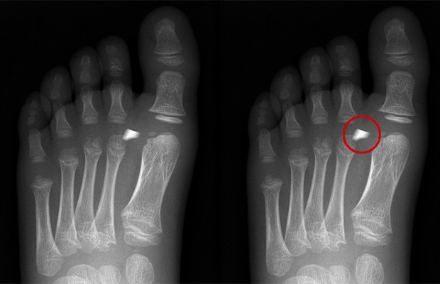
5. Check the Base of Fifth.
The metatarsal tuberosity is where the majority of fractures are found here.Peroneus brevis attaches here, and an inversion injury can result in a fracture.
6. Check for Lisfranc Injuries
On the DP view, the second metatarsal should line up with the intermediate cuneiform, and on the oblique view, the third metatarsal should line up with the lateral cuneiform. The second metatarsal and the cuneiforms are joined by the Lisfranc ligament. This ligament is crucial and should not be overlooked because damage to it results in an unstable foot.
7. Consider Stress Fractures
The second or third metatarsals are where these typically develop. They may occasionally only have callous formation or may not be visible on a plain x-ray, necessitating additional imaging (e.g. MRI) to diagnose.
Read More: Bump On Top Of Foot
How Do I Prepare for a Foot X-ray?
There isn’t much preparation needed for foot X-rays. You must take off any clothing, shoes, jewelry, or metal objects that could obstruct a clear X-ray image before the test.
Additionally, it’s crucial that you let your radiologic technologist know if you’re pregnant or think you might be. Radiation exposure to your developing child (fetus) is a possibility. If you need the X-ray, your doctor will make that determination. If an urgent situation necessitates taking an X-ray, safety measures will be taken to reduce your baby’s exposure to radiation.
The technician will go over the procedure with you before your X-ray. Before beginning the procedure, they will make sure you are prepared by responding to any questions you may have.
What Can I Expect During a Foot X-ray?
Your foot will be scanned by a radiologic technician in an X-ray room. This could happen in a hospital radiology department or in the office of your healthcare provider. A lead apron may be placed on your lap once you’re in the X-ray room by the technologist to shield your reproductive organs from radiation. If you are or might be pregnant, this is especially crucial. Although it might be a little chilly in the X-ray room, the entire process shouldn’t take more than 15 minutes.
Your leg will be placed on the X-ray table by the technician. The technologist may then secure your leg or foot with positioning equipment, like sandbags or pillows, to prevent movement. During the X-ray, it’s critical to maintain complete stillness because movement can blur the X-ray images.
Under the X-ray table will be a digital recording plate or an X-ray film holder, depending on the situation. The X-ray machine will then be turned on after they enter a special room or hide behind a wall. In order to obtain images from various angles, your technologist may move your foot several times. To make sure they get all the views, three separate images are typically taken. One image from the front, one from the side, and one at a 45-degree angle between the front and side views will all be taken by the technologist. Describe any pain you feel to your technologist. Throughout the test, they will help you and do everything in their power to make you feel at ease.
What Can I Expect After a Foot X-ray?
After the X-ray of your foot, the radiologic technologist will likely ask you to wait for a few minutes while they review the images. Before sending you on your way, they want to make sure the images are clear. They will immediately retake any blurry photos.
The images will then be examined by a physician known as a radiologist. Radiologists are educated in analyzing and interpreting X-ray images. The radiologist will send your healthcare provider the findings after reviewing them. Your healthcare provider will discuss the findings with you and choose the best course of action for your condition. A foot and ankle surgeon who has received specialized training in foot and ankle conditions may be recommended to you.
You might need to go back for a follow-up X-ray if your doctor or other medical professional requests more views of your foot or ankle. You might need to go back to monitor your health and keep an eye out for any changes that develop over time.
What Are the Risks of a Foot X-ray?
X-rays give your doctor a quick and easy way to identify any potential health issues with your foot or feet. A foot X-ray exposes you to a very small amount of radiation, and that radiation directly passes through you. Additionally, side effects from X-rays are extremely rare.
Pregnant women are somewhat more likely to experience radiation exposure problems. If you think you might be pregnant or are pregnant, you should always let your radiologic technologist know. To shield your reproductive system from radiation, you could put on a lead apron. Children are also somewhat more at risk. On children, lower radiation doses can be used.
Radiation overexposure carries a small risk of cancer. Healthcare professionals concur, though, that the advantage of a correct diagnosis outweighs the risk of radiation exposure. Ask your technician if you have any questions about the radiation exposure you’ll be receiving.
Also Check: What Is The Bunion Surgery Recovery Time?
Why Would I Need a Foot X-ray?
An X-ray of your foot may be taken to check for any indications of a foot or ankle injury, particularly following an accident. To rule out any underlying medical issues in your feet or ankles, your doctor may order a foot X-ray. These conditions may include:
- Your feet and ankles may have broken bones (fractures).
- Dislocated joints.
- Bunions and other joint deformities like hallux valgus.
- Arthritis in the foot and ankle is an example of a degenerative condition.
- Cysts.
- Bone infections.
- Bone cancer.
A foot X-ray can assist your healthcare provider in determining the cause of any unexplained swelling, pain, or tenderness in your foot or ankle.
After a broken ankle bone has been repaired, foot X-rays are frequently performed. Your doctor will be able to tell from the X-ray whether the bone was properly set. In order for bones to heal properly, they must be properly aligned.
FAQs
When Should You Get An X-ray for Foot Pain?
If you experience tenderness on the navicular bone (the bone on the inside part of your foot) by itself or in conjunction with either or both of the first two signs, then you should get yourself in for a foot X-ray.
What Can X-ray of Foot Show?
Using a foot X-ray, doctors can determine what is causing any pain, tenderness, swelling, or deformities. It can show broken bones or dislocated joints. An X-ray can be used to determine whether a broken bone has healed properly and whether the bones are in the correct alignment after being set.
How Many Types of Foot X-rays Are There?
There are two views in foot x-rays DP (dorsal-plantar) and oblique. Ideally, if your patient is able to handle it, both should be performed while bearing weight.
Who Performs a Foot X-ray?
Your foot will be radiographed by a radiologic technologist, or X-ray technician. The radiation protection, patient care, radiation exposure, radiographic positioning, and radiographic techniques that these specially trained healthcare professionals have studied.
Takeaway
When taken during an emergency, X-ray results are frequently available right away. Your radiologist will typically send the results to your doctor within one to two days if the situation is not urgent.
Your healthcare professional will then go over the results and treatment options with you.
Tags: Foot X Rays